
Anatomy
Vertucci canal pathways: read the route before you shape
A visual guide to the eight classic canal configurations—and the clinical questions each pathway should prompt.
Clinical frame
The number is a pathway—not just a canal count.
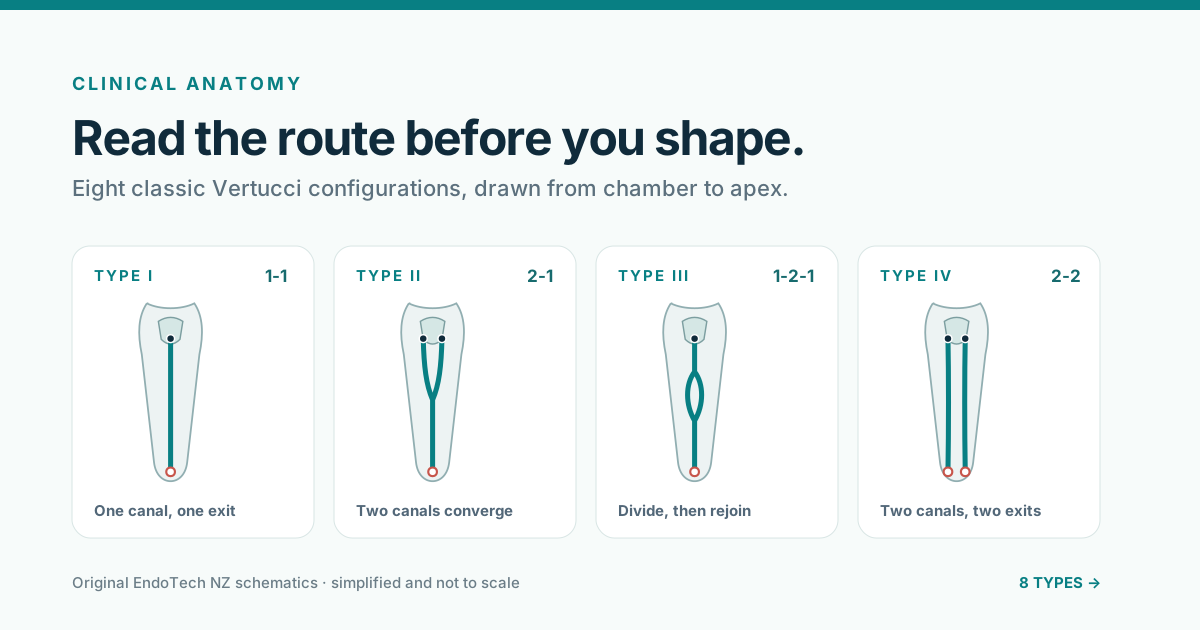
Vertucci classification describes how canals begin coronally, divide or merge through the root, and finish apically. Read the sequence from chamber to apex: 2-1, for example, means two distinct canals converge into one exit.
Critical to have case-specific diagnosis, imaging, magnification, or clinical judgement.
Anatomy map
The eight classic configurations.
One canal, one exit
Two canals converge
Divide, then rejoin
Two canals, two exits
One canal divides
Merge, then redivide
Divide, join, divide
Three canals, three exits
Original EndoTech NZ schematics. Simplified, not to scale, and intended for education—not anatomical prediction.
Chairside application
Classification should guide you to – 1, 2, 3.
Naming the likely pathway is useful only when it changes what you look for and how deliberately you confirm it.
Find the entry pattern
Count distinct coronal canal entries and look for developmental grooves that may connect or conceal them.
Verify every transition
Use angled radiographs, magnification, tactile feedback, an apex locator, and CBCT when clinically indicated to test the pathway you think is present.
Preserve the pathway
Establish a reproducible glide path and shape with enough control to respect divisions, curves, convergence, and separate exits.
human permanent teeth in Vertucci's landmark cleared-tooth investigation.
Primary source
Root canal anatomy of the human permanent teeth.
Vertucci FJ. Oral Surgery, Oral Medicine, and Oral Pathology. 1984;58(5):589-599.
View the PubMed record